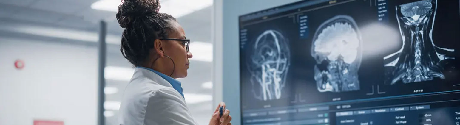
The past year has solidified the role of medical imaging not just as a diagnostic tool, but as an integral part of precision medicine and therapy. The key theme across all modalities—from X-ray to MRI—is the move toward innovative, fast, and personalized imaging protocols.
I. AI-Driven Automation and Augmentation
Artificial Intelligence is the single most transformative force in modern radiology, driving dozens of micro-advancements that collectively enhance the entire workflow.
1. AI-Powered Image Reconstruction (Deep Learning Reconstruction)
This is a significant breakthrough across all modalities (CT, MRI, PET). Deep learning algorithms are used to reconstruct high-quality images from less data.
Impact: Dramatically reduces scan times (improving patient comfort and throughput) and allows for a significant reduction in radiation dose (for CT) or contrast agent use, while maintaining or even improving image resolution.
2. Automated Quantification and Segmentation
AI models now routinely perform tedious, time-consuming tasks:
Organ and Lesion Segmentation: Automatically outlining tumors, organs, and structures (e.g., measuring liver volume or segmenting cardiac chambers), which is critical for surgical planning and treatment monitoring.
Quantitative Metrics: Generating automated measurements, such as tracking tumor growth or calculating cardiac ejection fraction, leading to more objective and reproducible reports.
3. Workflow Prioritization and Triage
AI systems are now integrated into Picture Archiving and Communication Systems (PACS) to analyze incoming studies and flag critical findings (e.g., intracranial hemorrhage, pulmonary embolism) for the radiologist to review immediately.
Impact: Improves response time for time-sensitive, life-threatening conditions.
4. Generative AI for Reporting
The initial adoption of Large Language Models (LLMs) is beginning to draft routine sections of radiology reports, summarize complex patient histories, and even help in communicating findings to patients in plain language.
II. Modality-Specific Hardware Breakthroughs
Next-generation hardware is pushing the limits of image quality and functional analysis.
5. Photon-Counting CT (PCCT)
PCCT is replacing conventional CT detectors.
Mechanism: Instead of measuring the total energy of an X-ray beam, PCCT counts the number of individual photons and measures their specific energy levels.
Impact: Unprecedented spectral imaging (color CT), much higher resolution, improved contrast-to-noise ratio, and the ability to differentiate materials with far greater sensitivity than traditional CT.
6. Ultra-High Field MRI (7T and beyond)
The clinical availability of 7-Tesla (7T) MRI systems is expanding.
Impact: Allows non-invasive imaging with exquisite detail of subtle brain structures, small-vessel disease, and high-resolution cartilage and musculoskeletal structures, enhancing the diagnosis of complex neurological disorders and joint conditions.
7. Mobile and Portable Imaging (MRI and POCUS)
Advancements have made high-quality imaging more accessible:
Helium-Free/Low-Field MRI: Smaller, easier-to-install MRI systems requiring no cryogen infrastructure, allowing high-quality scans to be deployed in outpatient centers or even remote locations.
Point-of-Care Ultrasound (POCUS): Pocket-sized, app-driven ultrasound devices are becoming standard equipment for physicians across specialities, transforming real-time triage and procedure guidance.
8. Total-Body PET (TB-PET)
New scanners with extremely long axial fields of view (up to two meters) can image the entire body simultaneously.
Impact: Significantly reduces scan time (down to seconds) and allows for dose reduction, making it ideal for tracking circulating immune cells, whole-body cancer staging, and pediatric imaging.
III. Precision and Functional Imaging
The ability to image function—not just structure—is becoming standard practice.
9. Theragnostic-Guided Imaging
The rise of theragnostic (the combination of therapy and diagnostics) is a major driver. New radiopharmaceuticals, such as those targeting Prostate-Specific Membrane Antigen (PSMA) for prostate cancer, require particular PET and SPECT imaging for patient selection and monitoring of treatment response.
10. AI-Enhanced Functional MRI (fMRI) and Diffusion Tensor Imaging (DTI)
AI is making complex functional studies (e.g., mapping brain activity or white matter tracts) more robust, faster to acquire, and easier to interpret, directly assisting in pre-surgical planning for brain and spine procedures.
11. Multi-Modal and Fused Imaging Refinement
The routine, seamless fusion of images from different modalities (e.g., PET-CT, PET-MRI) is becoming a core clinical tool, leveraging AI to automatically align and register images to provide a complete picture of anatomy and metabolism in one view.
IV. The Digital & Interventional Environment
Advances in how images are stored, accessed, and used during procedures are boosting efficiency and safety.
12. Enterprise Imaging and Cloud Solutions
A shift from localized PACS systems to cloud-based Enterprise Imaging Platforms allows clinicians across a health system (and even outside partners) to access images and reports securely and instantly, enabling remote consultation and faster diagnosis.
13. Advanced Dose Reduction Protocols
Ongoing software and hardware advancements continue to reduce the ionizing radiation dose delivered by CT and X-ray systems, often using iterative reconstruction algorithms far beyond what was possible just a few years ago.
14. Augmented Reality (AR) in Interventional Radiology
Interventional radiologists are using AR and Virtual Reality (VR) to superimpose 3D anatomical models onto the patient during minimally invasive procedures.
Impact: Allows for enhanced guidance of needles, catheters, and ablation probes, improving targeting accuracy and reducing risk.
Conclusion
While a list of individual clinical advances could fill volumes, the major thrust in medical imaging is clear: intelligence, integration, and accessibility. AI is no longer a research tool but a fundamental component of the scanner and the reporting process, guaranteeing higher precision and efficiency for every patient. The focus is now on making these advanced, functional imaging techniques available to all, ensuring that the proper diagnosis is reached faster and treatment is optimally planned.